2021年度第4回ExCELLSセミナーを開催いたします。
日時
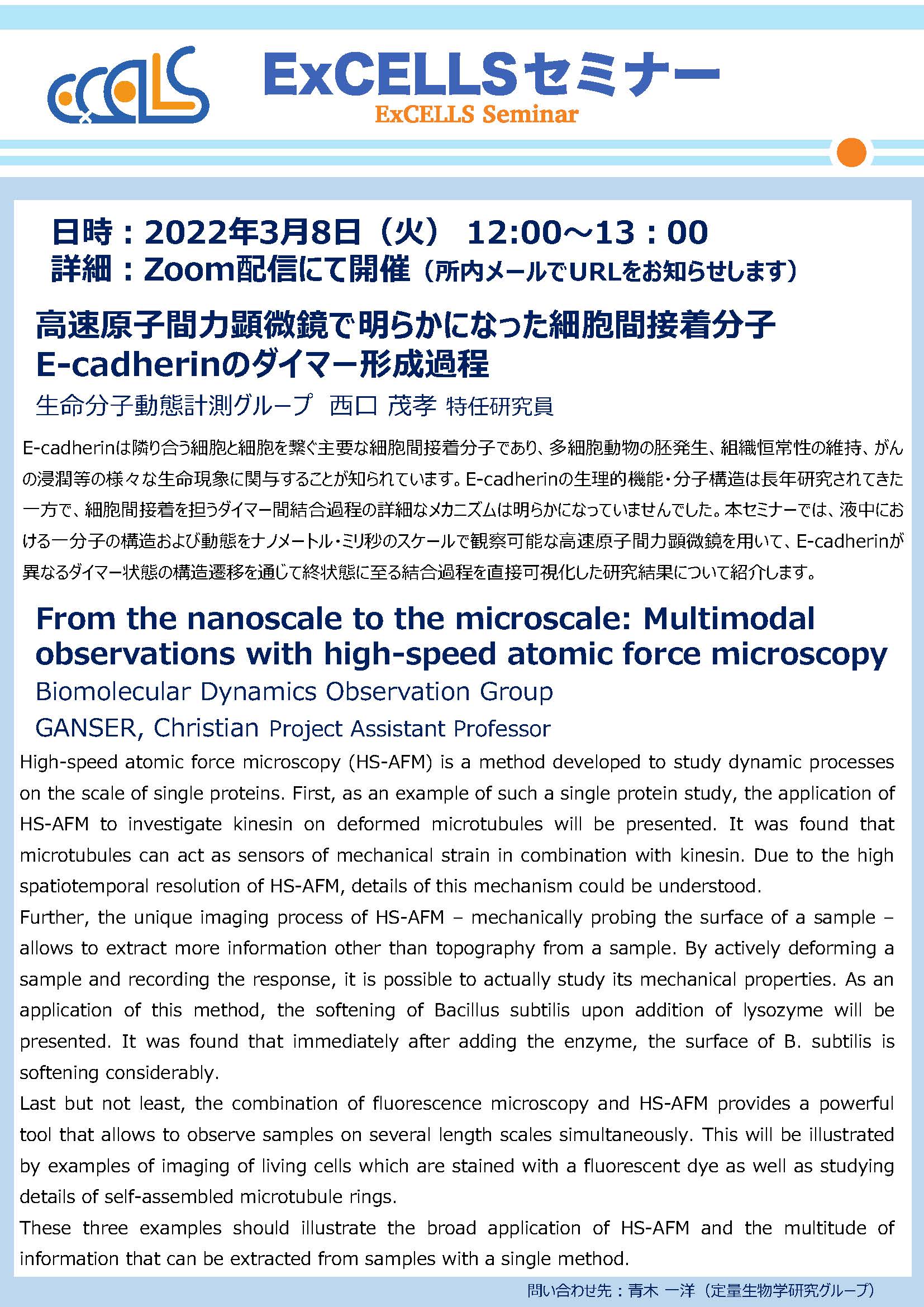
2022年3月8日(火) 12:00〜13:00
場所
Zoomオンライン
演者
- 西口 茂孝 特任研究員
(創成研究領域 生命分子動態計測グループ) - GANSER, Christian Project Assistant Professor
(Biomolecular Dynamics Observation Group, Department of Creative Research)
題目
高速原子間力顕微鏡で明らかになった細胞間接着分子E-cadherinのダイマー形成過程
(西口 茂孝 特任研究員)
From the nanoscale to the microscale: Multimodal observations with high-speed atomic force microscopy
(GANSER, Christian Project Assistant Professor)
要旨
- 高速原子間力顕微鏡で明らかになった細胞間接着分子E-cadherinのダイマー形成過程
E-cadherinは隣り合う細胞と細胞を繋ぐ主要な細胞間接着分子であり、多細胞動物の胚発生、組織恒常性の維持、がんの浸潤等の様々な生命現象に関与することが知られています。E-cadherinの生理的機能・分子構造は長年研究されてきた一方で、細胞間接着を担うダイマー間結合過程の詳細なメカニズムは明らかになっていませんでした。本セミナーでは、液中における一分子の構造および動態をナノメートル・ミリ秒のスケールで観察可能な高速原子間力顕微鏡を用いて、E-cadherinが異なるダイマー状態の構造遷移を通じて終状態に至る結合過程を直接可視化した研究結果について紹介します。
- From the nanoscale to the microscale: Multimodal observations with high-speed atomic force microscopy
High-speed atomic force microscopy (HS-AFM) is a method developed to study dynamic processes on the scale of single proteins. First, as an example of such a single protein study, the application of HS-AFM to investigate kinesin on deformed microtubules will be presented. It was found that microtubules can act as sensors of mechanical strain in combination with kinesin. Due to the high spatiotemporal resolution of HS-AFM, details of this mechanism could be understood.
Further, the unique imaging process of HS-AFM – mechanically probing the surface of a sample – allows to extract more information other than topography from a sample. By actively deforming a sample and recording the response, it is possible to actually study its mechanical properties. As an application of this method, the softening of Bacillus subtilis upon addition of lysozyme will be presented. It was found that immediately after adding the enzyme, the surface of B. subtilis is softening considerably.
Last but not least, the combination of fluorescence microscopy and HS-AFM provides a powerful tool that allows to observe samples on several length scales simultaneously. This will be illustrated by examples of imaging of living cells which are stained with a fluorescent dye as well as studying details of self-assembled microtubule rings.
These three examples should illustrate the broad application of HS-AFM and the multitude of information that can be extracted from samples with a single method.
ポスター
お問い合わせ先
青木 一洋(定量生物学研究グループ)
当日の様子