
Six structures exhibited by the rotating sodium ion pump were reconstructed in 3D using cryo-electron microscopy. This analysis revealed that (1) the rotor exhibits non-uniform rotation behavior due to partial structural interference with the stator component, and (2) the rotor interacts with one edge of the large ion transport ring causing it to rotate. The study showed a unique molecular mechanism of the rotary sodium ion pump.
The results will be published on July 28 in “Communications Biology”.
“In previous single-molecule imaging experiments, it was predicted that the three main pausing points of the rotor would be 120 degrees apart, and each sub-pause would be 40 degrees ahead (80 degrees back) from each main pause, but there was no structural evidence. It was also a mystery how the thin shaft (rotor) that connects the power unit rotates the sodium ion transport ring, which is larger than this unit.” said corresponding author Kazuyoshi Murata, project professor, Exploratory Research Center on Life and Living Systems (ExCELLS) and National Institute for Physiological Sciences (NIPS) in Japan. “The rotor turns the large ion transport ring as if it were stirring with uneven rotation behavior.”
- Research background
Almost all living organisms, from bacteria to humans, have the rotary enzyme ATP synthase, in their cell membranes. These all have a similar structure in which an extramembrane part for decomposing or synthesizing the compound ATP and an intramembrane part for passing ions across the membrane are connected by a thin shaft (rotor) to provide dual functionality. ATP synthase can use both the energy of the passive flow of ions across the membrane to synthesize ATP, while ions can be actively transported across the membrane by decomposing ATP.
The rotary sodium ion pump (EhV-ATPase) of Enterococcus hirae (Fig. 1) decomposes ATP in the extramembrane power unit (V1) and rotates the rotor at its center, which rotates the sodium ion transport ring (c-ring) of the intramembrane pump unit (Vo). This transports sodium ions from the inside to the outside of the cell. The power unit consists of three symmetrically arranged ATP degradation domains (consisting of an A/B heterodimer), which are known to rotate the rotor by 120 degrees through sequential degradation of ATP. Our research group previously labeled this power unit with a gold nanoparticle and performed single-molecule imaging experiments, and found that sub-pauses (State 1′ to 3′) exist at each position 40 degrees ahead (80 degrees back) of the main pauses (State 1 to 3), and identified six “pause” states. Also, EhV-ATPase has a large c-ring, like V-ATPase in higher organisms, but there is no other protein subunit that connects the rotator straight to the center of the c-ring, as seen in higher organisms. Therefore, in EhV-ATPase, (1) what the six structures including the sub-pause are, and (2) how the rotator rotates the large c-ring remained a mystery.
- Research result
In this study, in order to answer these questions, we induced rotation by adding ATP and sodium ions to detergent-solubilized EhV-ATPase, rapidly froze the sample, and collected many images using cryo-electron microscopy. After classifying the obtained images and reconstructing them in 3D, we succeeded in reconstructing a total of 6 structures consisted of 3 main pauses and 3 sub-pauses for the first time (Fig. 2A).
As described above, the rotor rotation of EhV-ATPase was supposed to stop at the main pausing points (States 1 to 3) separated 120°. This was true in States 1 and 2, but it was found that there was a -10° deviation in the rotor angle in State 3 (Fig. 2A). In addition, at the sub-pauses (State 1′ to 3′), large angle deviations of +13° (State 1′), +32° (State 2′), and +28° (State 3′) from those expected were observed. Then, it was found that the rotor edge deviated from the center of the c-ring and interacted to one edge of the c-ring, causing the c-ring to stir and rotate (Fig. 2B). This rotational angle deviation is particularly pronounced between States 2 and 3′, and it is thought that these are caused by structural interference between the subunit “d” of the rotor edge and the adjacent stator subunit “a” (Fig. 2C).
In the power unit V1, we were able to partially confirm changes in the bound and decomposed states of ATP during rotational motion (Fig. 3A). Based on previous studies, it is believed that the three symmetrically arranged ATP binding domains sequentially exhibit three states: “Closed” with ATP bound, “Semi-closed” with ATP decomposed into ADP and phosphate (Pi), and “Open” with no ATP or ADP bound. By applying these states to the obtained structural map, we were able to confirm the conformation of the ATP binding structure at the three main pauses. On the other hand, at the three sub-pauses, except for State 2′, we obtained structures that are considered to be in a state in which all the ATP binding pockets are filled with ATP or ADP before ADP is released and the domain becomes “Open” (Fig. 3). As mentioned above, in State 2’, structural interference between the rotor and the stator was thought to cause the ATP binding domain to proceed to the same state as in State 3 and stop (Fig. 3B).
- Significance of results and future perspective
In this study, by actively inducing rotation and by analyzing the structures during rotor rotation, we were able to visualize six structures (three main pauses and three sub-pauses) of EhV-ATPase. This result clarified that the EhV-ATPase transports sodium ions by rotating a large c-ring and revealed that structural interference of the rotor with a part of the stator (subunit “a”) caused a deviation in the rotation angle. These findings suggest an adaptive process from a simple to a more complex structures in the evolution of the nearly ubiquitous rotary ion pumps, and have provided much structural information for the development of inhibitors and functional modification of enzymes. Based on this result, it is expected that drug discovery and development targeting this ion pump will progress greatly in the future.
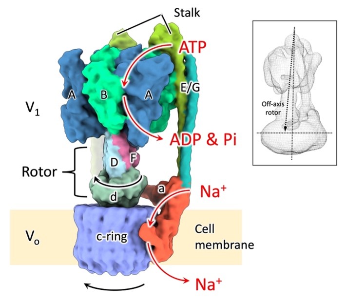 Figure 1. Structure of the rotary sodium ion pump EhV-ATPase. It consists of V1, which hydrolyzes ATP outside the cell membrane and rotates the central rotor subunits “D/F/d”, and Vo, which rotates the c-ring inside the cell membrane to transport sodium ions (Na+) from the inside to the outside of the cell. V1 is composed of seven protein subunits, “A, B, D, E, F, G, and d”, and Vo is composed of two protein subunits, “a and c”. V1 and Vo are connected by a subunit “E/G” called “stator” to fix the V1 and Vo domains. Previous studies have shown that the rotor tilts and connects with the c-ring (inset), but how the rotor rotates the c-ring remained a mystery.
Image credit: Kazuyoshi Murata
Image usage restrictions: Creative Commons licence CC BY 4.0
 Figure 2. Six structures exhibited by EhV-ATPase. States 1 to 3 indicate main pauses and States 1′ to 3′ indicate sub-pauses. The main pauses are structures in which the rotor rotates by 120 degrees, but in State 3, the angle deviated -10 degrees. In addition, the sub-pauses deviated +13° in State1′, +32° in State2′, and +28° in State3′. (A) Overall structure of EhV-ATPase. (B) Positional relationship between the rotor terminal subunit “d” and the c-ring. Subunit “d” (red “+” indicates the position of the centroid) is in contact with the c-ring at a position distant from the center of the c-ring (black “+”). As a result, the rotor stirs the c-ring as indicated by the yellow line in (A). (C) Structural interference between rotor-end subunit “d” and stator subunit “a”.
Image credit: Kazuyoshi Murata
Image usage restrictions: Creative Commons licence CC BY 4.0
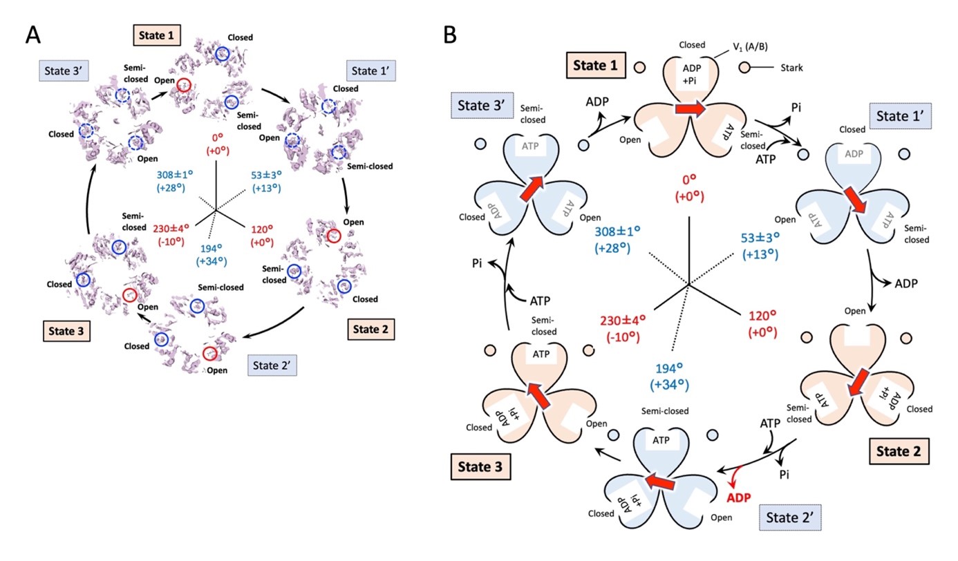 Fig. 3. ATP decomposition and rotor rotation. Structural changes in the ATP binding domain of V1 in the six structures of EhV-ATPase (A) and a schematic diagram of the relationship between the ATP decomposing reaction and the rotational motion of the rotor (B).
Image credit: Kazuyoshi Murata
Image usage restrictions: Creative Commons licence CC BY 4.0
- Published paper
Journal name: Communications Biology
Paper title: Six states of Enterococcus hirae V-type ATPase reveals non-uniform rotor rotation during turnover
Authors: Raymond N. Burton Smith, Chihong Song, Hiroshi Ueno, Takeshi Murata, Ryota Iino, Kazuyoshi Murata* (*corresponding author)
Posting date: 2023 UK time Friday July 28th 10am
DOIs: 10.1038/s42003-023-05110-8
Contact
Media contact :
Strategic Research Administration Office
Exploratory Research Center on Life and Living Systems (ExCELLS)
National Institutes of Natural Sciences
E-Mail: press_at_excells.orion.ac.jp
(Please replace the “_at_” with @)
Expert contacts :
MURATA, Kazuyoshi
Exploratory Research Center on Life and Living Systems (ExCELLS)
National Institute for Physiological Sciences (NIPS)
Natural Institutes of Natural Sciences, Okazaki
E-Mail: kazum_at_nips.ac.jp
(Please replace the “_at_” with @)