
ExCELLS promotes collaborative research with researchers from other institutes and universities. Various researchers can use ExCELLS equipment to perform their own research through joint research projects along with ExCELLS researchers.
ExCELLS welcomes an application of from researchers who want to use ExCELLS’s equipment.
Click here for application guidelines. Click here for application information.
If you have any questions, please feel free to the contact desk (collabo_at_excells.orion.ac.jp).
*Please replace the “_at_” with @
The following is an introduction for equipment available in ExCELLS joint research.
ExCELLS Equipments
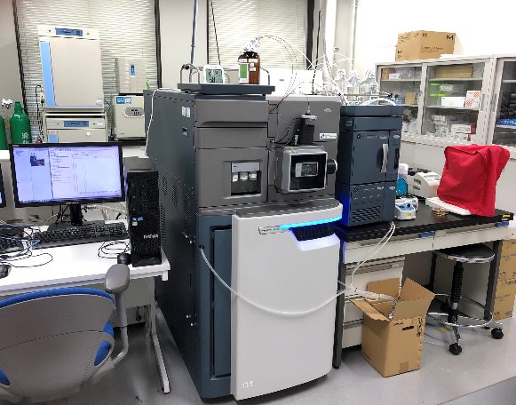
System of Cryo-electron microscopy
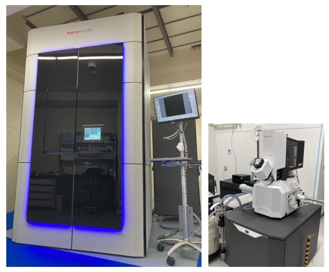
This system consists of a 300kV cryo-EM (TITAN Krios G4) and a cryo FIB-SEM (Aquilos2). TITAN Krios G4 can perform atomic resolution single particle analysis of proteins, electron tomography, and MicroED. Aquilos2 can generate frozen cell sections for in situ electron tomography, in addition to Slice & View observations of frozen samples.
800 MHz NMR spectrometer
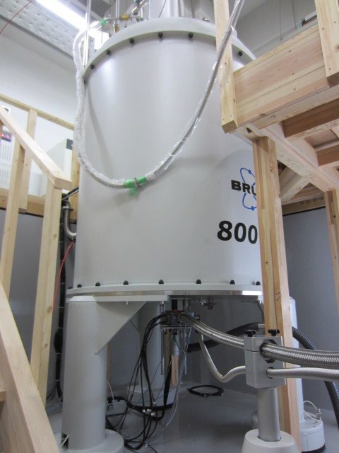
This NMR spectrometer (Bruker 800 MHz NMR AVANCE NEO 800US equipped with a cryogenic probe) is capable of 1H-13C-15N triple resonance measurements of biomacromolecules at high sensitivity and high resolution for characterizing their structural dynamics and interactions at atomic resolution.
Combined system of high-speed atomic force microscopy and fluorescence microscopy
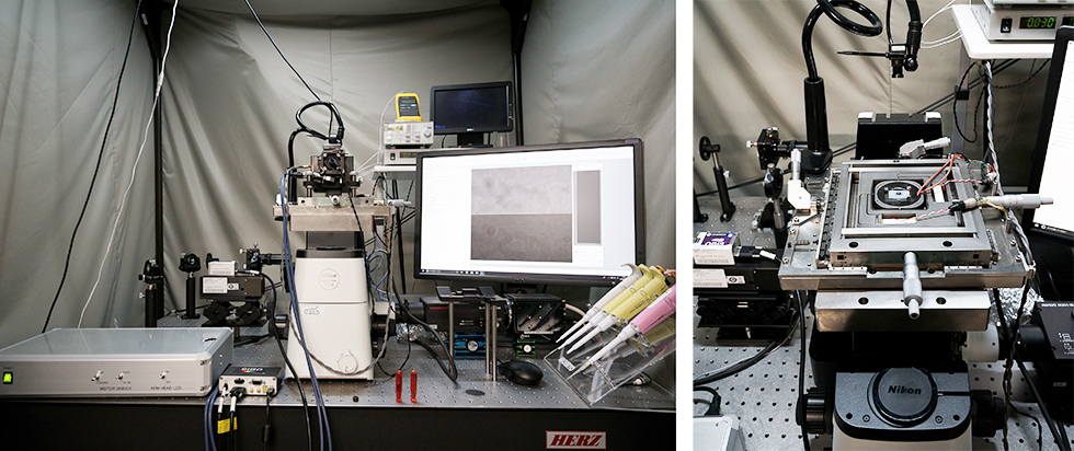
The combined high-speed atomic force microscopy (HS-AFM) and fluorescence microscopy can visualize the dynamic phenomena of various biological samples from proteins to living cells in real time. This equipment can visualize biomolecular behaviors simultaneously with HS-AFM and fluorescence microscopy.
Mass spectrometers for native MS
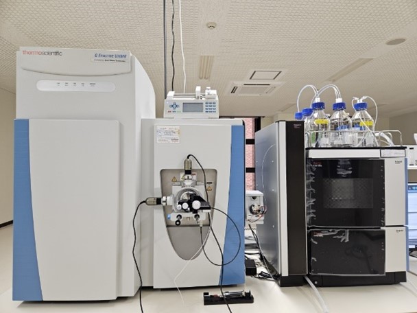
ExCELLS has two different mass spectrometers for native MS. (Left) One is a Q Exactive UHMR Hybrid Quadrupole Orbitrap Mass Spectrometer (Thermo Fisher Scientific), which is a Q-Orbitrap mass spectrometer with the features of high resolution and wide range of m/z. Due to these strong features, biomolecular complexes with high heterogeneities and supramolecular assemblies such as virus capsids can be analyzed. (Right) The other is a SYNAPT G2-Si HDMS system (Waters), which is a Q-TOF mass spectrometer with ion mobility mass spectrometry (IM-MS) mode. LC-MS/MS is also available.
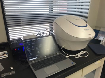
Mass Photometer
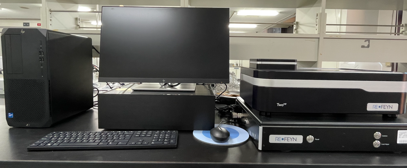
Mass photometry measures mass at the single molecule level, providing insights into the composition and function of even complex samples and molecular mechanisms. This unique technology can be used to detect and characterise proteins, nucleic acids, lipids and/or sugars, providing information on structure (oligomeric state, modification), homogeneity, and function (quantifying interactions) – all in a matter of minutes, and using tiny amounts of sample. The Refeyn TwoMP enables measurement of molecular mass across a wide mass range, from 30 kDa up to 5 MDa.
Biomolecular interaction analysis system
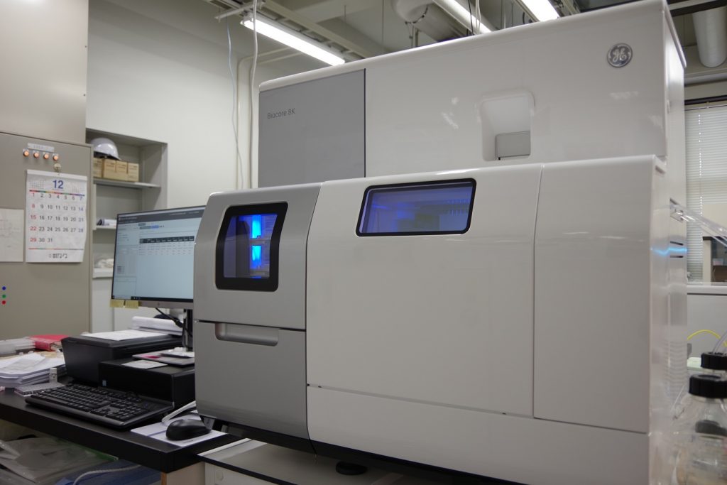
This system detects intermolecular interaction by surface plasmon resonance, enabling comprehensive, high-throughput analysis to obtain quantitative kinetic and affinity data.
Dynamic Light Scattering instrument
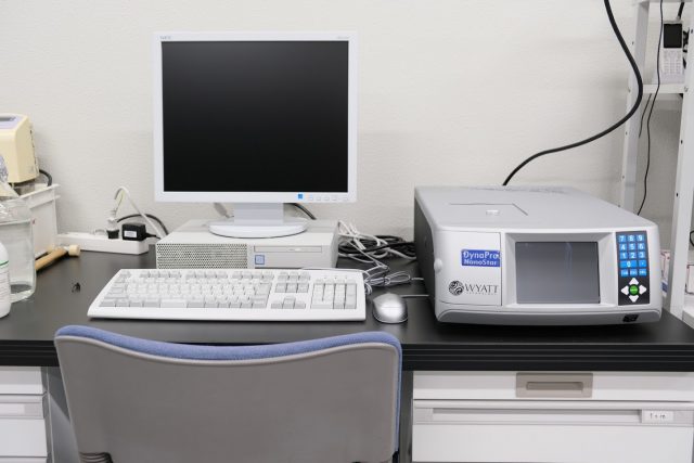
DynaPro Nanostar (Wyatt technology) is a device to measure the size distribution of biomolecules such as proteins and nano particles using Dynamic Light Scattering (DLS). We can also measure molecular weight of biomolecules and viscosity of solutions. For measurements, only several µL of sample is required and a wide range of temperature (−15℃ ~ 150℃) is available.
Temperature-controlled microvolume spectrophotometer
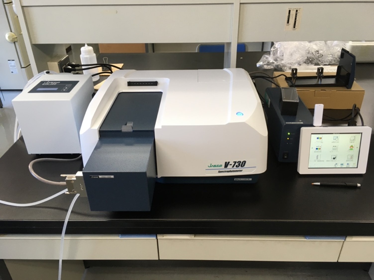
This instrument, the V-730 spectrophotometer by Japan Spectro Co., Ltd. is a spectrophotometer capable of measuring absorbance from 190 to 1100 nm. Notably, it is equipped with an 8-cell microcell block and a Peltier temperature controller. These components enable precise sample temperature control over a wide range from 0 to 100℃, and can handle up to eight micro-volume samples (10μl each) simultaneously. This functionality enables the automatic measurement of multiple samples with different conditions such as concentration, while varying the temperature. Additionally, it is equipped with a direct sensor for sample temperature measurement, enabling accurate monitoring of sample temperature. These features make it possible to study phenomena affected by temperature changes such as liquid-liquid phase separation, enzyme reactions, and the thermal stability of nucleic acids.
Fluorescence and Absorbance Spectrometer
This system is capable of high-speed spectral measurement of absorbance and fluorescence from UV to near-infrared with time intervals of less than 100 ms. Since the Peltier temperature controller allows various temperature controls from ultra-low to high temperatures, it is useful for obtaining the temperature characteristics of fluorescent proteins at various temperatures. In addition, this device can measure absorbance and fluorescence simultaneously and is expected to be utilized for component analysis by excitation fluorescence matrix and FRET analysis, etc.
Super-resolution microscopy systems
Multifunctional super-resolution confocal fluorescence microscope
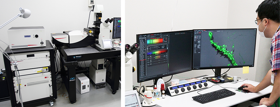
This multifunctional confocal microscope enables super-resolution, fluorescence lifetime measurement, and fluorescence correlation microscopy.
- Super-resolution microscopy
– STED (stimulated emission depletion) can resolve below 50 nm.
– An easy-to-use deconvolution program “Lightning” archive ~120 nm resolution. - Fluorescence lifetime imaging microscopy (FLIM)
FALCON (FAst Lifetime CONtrast) enables much faster fluorescent lifetime imaging compared to conventional systems, that is applicable for detection of microenvironmental changes using molecular biosensors, and for separation of multiple fluorophores by their lifetime difference. - Fluorescence Correlation Spectroscopy (FCS)
FCS is a correlation analysis of fluctuation of the fluorescence intensity that provides quantitative parameters of the physics such as concentration, diffusion coefficient, etc. FCS combined with STED and FALCON enables finer quantification.
Two-photon STED microscope
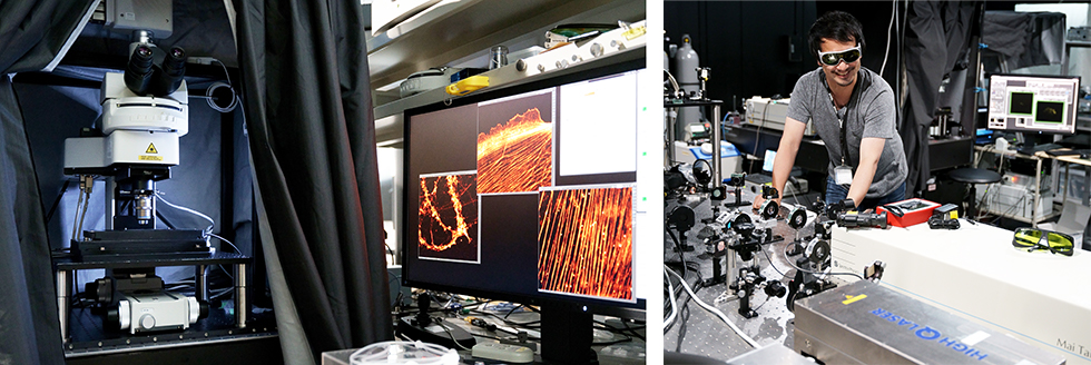
It enables super-resolution microscopic observation of two-photon excited fluorescence and/or fluorescence lifetime imaging.
Lattice Light-sheet Microscope
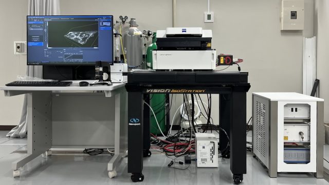
This microscope, ZEISS Lattice Lightsheet 7 is a combination of light-sheet microscopy, lattice-like structured illumination, and integrated incubation system. It enables volumetric imaging of living cells and tissues with high-speed (~3 vol/sec), low photo-damages, and spatial resolution as high as confocal microscopes.
High-speed live imaging system
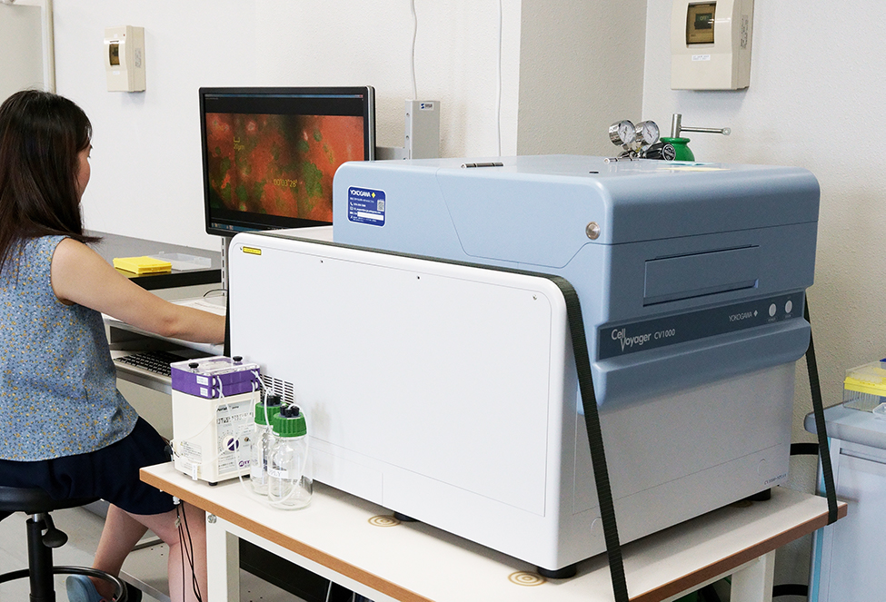
Spinning disk confocal microscopy equipped with cell culture device. Three lasers (488, 560, 640 nm) are equipped, and long-term live imaging (~ 1week) can be performed.
Total internal reflection fluorescence (TIRF) microscope
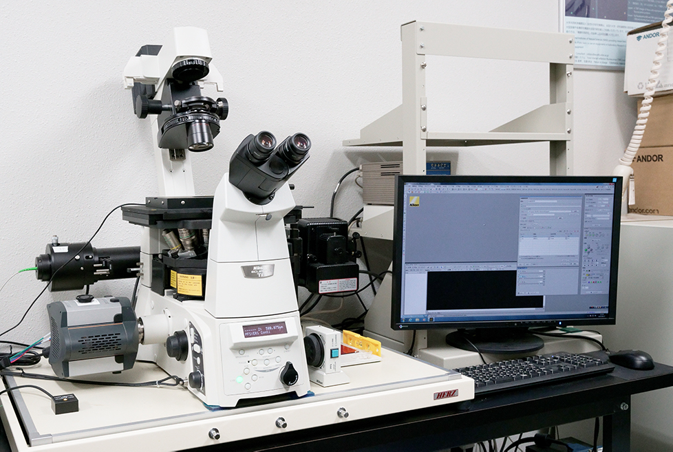
Total internal reflection fluorescence (TIRF) lasers (405, 488, 561 nm) are equipped with electric inverted microscope using EM-CCD (Andor) detector. Single molecule measurement and HILO imaging can be performed using 100X TIRF objective lens.
4-Dimensional Tissue Imaging System
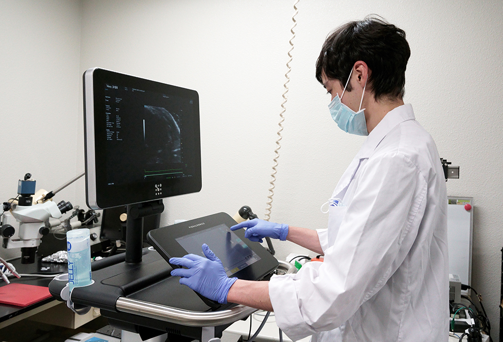
This equipment enables us to reconstruct tissue dynamics at the 4-dimensional level by measuring structural and functional changes of tissue and organ of your interest during the developmental or pathological processes using small animals (e.g., mouse and rat).
Cell sorting and measurement system
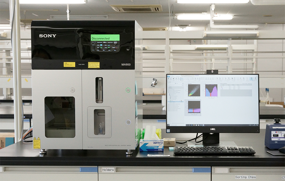
MA900 Multi-Application Cell Sorter allows detection of up to 12 fluorescence at the single-cell level using fluorescent proteins and/or antibodies labeled with fluorescent dyes, and the gated target cells can be sorted for further analysis.
The cell sorter can automatically adjust various sorting settings (laser beam, optical axis adjustment, electrical timing adjustment for sorting, side stream adjustment, collection tube position adjustment). Four excitation lasers – 405 nm, 488 nm, 561 nm, 638 nm – are equipped, and 96 wells and 384 well plates are available for cell sorting.
Single-cell multi-omics analysis system
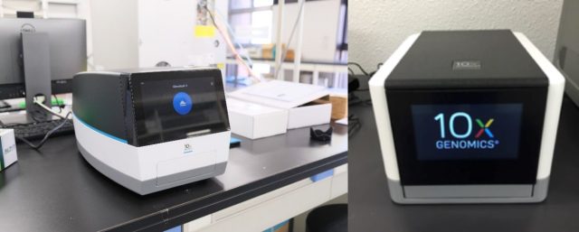
A single cell (or cell nucleus) is mixed with molecularly barcoded gel beads in a microdroplet to form tens and hundreds of thousands of droplets in the device (Chromium X & Chromium Controller system, 10x Genomics inc.). By applying the appropriate treatment within each microdroplet, multi-omics information such as gene expression, chromatin accessibility, and cell surface protein information within a single cell can be obtained by combining with NGS (Next Generation Sequencing). Up to 750,000 (Chromium X) and 80,000 (Chromium Controller) single cell information can be acquired in a single experiment.
Sophion QPatch Compact
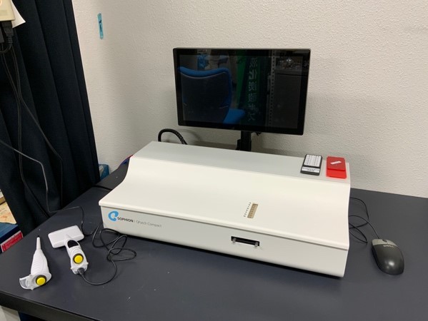
Sophion QPtach Compact is a semi-automatic patch-clamp system which allows easy 8 data acquisition as the same time for the scientists not familiar with electrophysiology. Using this system is especially suitable when a lot of data acquisition with a same experimental condition in a short time period or agonist/antagonist screening is needed. Original software made by Sophion supports the experiments and data analysis. Things you can do are cell preparation and pipetting.
Laser-Based quartz glass micropipette puller
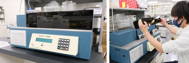
P-2000 micropipette puller (Sutter) is equipped with a CO2 laser-based heat source and can manufacture quartz glass pipettes. Quartz glass has extremely high physical strength and can achieve a tip diameter of 0.01 micron or less, which is not possible with ordinary glass. In addition, the CO2 laser is not easily affected by the external environment such as temperature and humidity, and has high reproducibility.
Chemiluminescence and fluorescence imaging systems
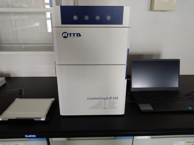
LuminoGraph II EM with F0.8 ultra-sensitive lens and ultra-low noise EM CCD camera allows to detect chemiluminescence and fluorescence signal from Western blot membranes and SDS-PAGE gels. Four LEDs (Blue, Green, Red, NIR) and white transilluminator are equipped.