
Residue-specific methyl signals enable practical structural evaluation of antibody drugs and biosimilars
Therapeutic antibodies are among the most widely used biologic medicines, yet detecting subtle structural differences in these complex proteins remains challenging. Researchers in Japan have established a nuclear magnetic resonance (NMR) strategy that reveals residue-specific structural features of antibodies without the need for isotope labeling.
The approach enables atomic-level evaluation of antibody structure, glycosylation, and molecular dynamics, providing a practical platform for quality assessment of biologic drugs and biosimilars.
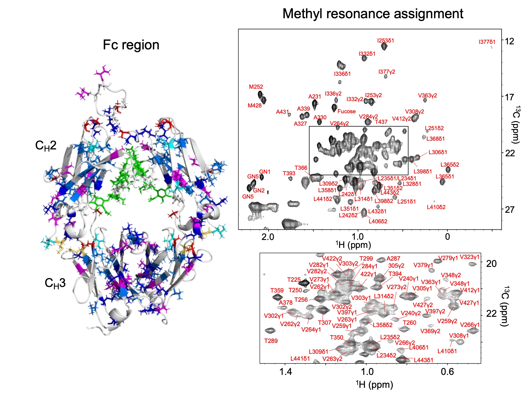
Map of NMR Signals from Methyl Groups in the Antibody Fc Region
Left: Residues containing methyl groups are shown on a three-dimensional model, color‑coded according to amino acid type.
Right: NMR signals of the antibody Fc region and their assignments.
Monoclonal antibodies are widely used to treat diseases ranging from cancer to autoimmune disorders. The safety and efficacy of these biologic drugs depend on maintaining their correct three-dimensional organization, known as their higher-order structure (HOS).
However, detecting subtle structural variations in antibodies remains a major analytical challenge. Conventional techniques such as circular dichroism or calorimetry provide valuable global information but generally lack residue-level resolution.
Nuclear magnetic resonance (NMR) spectroscopy is one of the few techniques capable of observing protein structure and dynamics in solution at atomic resolution. In recent years, NMR fingerprinting approaches have been increasingly used to evaluate higher-order structure in therapeutic antibodies. Yet most of these approaches detect overall spectral differences rather than providing detailed structural interpretation.
In a study published in the Journal of the American Chemical Society, researchers from the Exploratory Research Center on Life and Living Systems (ExCELLS) and collaborating institutions established site-specific assignments of methyl signals in the Fc region of human IgG1 antibodies.
The researchers combined amino-acid-selective isotope labeling, mutagenesis, and multidimensional NMR experiments to assign methyl resonances from multiple residues—including alanine, isoleucine, leucine, methionine, threonine, and valine—in the Fc domain.
These assignments allow methyl signals to function as residue-specific probes of antibody structure. Importantly, the same signals were detectable even in antibodies at natural isotopic abundance, meaning that isotope labeling is not required for structural analysis.
Using these probes, the researchers detected structural differences associated with glycosylation patterns in the Fc region, including variations in core fucosylation and terminal galactosylation—two glycan features known to influence antibody effector functions.
The study also revealed that certain methyl signals originate from highly dynamic regions of the antibody, including residues in the hinge region and receptor-binding interface. These signals act as sensitive reporters of local structural flexibility, providing insight into functionally important regions of the molecule.
Because the method can analyze antibodies directly in their unlabeled form, it opens new possibilities for monitoring structural integrity and subtle molecular variations in therapeutic antibodies. Such capabilities are increasingly important for ensuring the quality of biologic medicines and for demonstrating structural comparability in biosimilar development.
Quote
“Therapeutic antibodies are structurally complex molecules, and even subtle structural variations can influence their biological activity,” said Koichi Kato of the Exploratory Research Center on Life and Living Systems (ExCELLS), National Institutes of Natural Sciences.
“Our study shows that methyl-based NMR signals can serve as powerful structural probes even in unlabeled antibodies, providing a practical route to residue-level structural evaluation of antibody therapeutics.”
Related Research
This study complements a related paper published in Analytical Chemistry, in which the researchers developed an integrated NMR and LC–MS approach to characterize methionine oxidation in the IgG1 Fc region at residue-specific and stereochemical resolution.
That work revealed how oxidation at the conserved Fc residues Met252 and Met428 produces distinct R- and S-sulfoxide forms and perturbs local antibody structure, providing mechanistic insight into how oxidative modifications affect antibody stability and function.
Together, the two studies establish a comprehensive analytical framework for evaluating both higher-order structural features and chemical modifications of therapeutic antibodies.
Paper information
Saeko Yanaka, Yuuki Koseki, Yohei Miyanoiri, Toshio Yamazaki, Tsutomu Terauchi, Daichi Kaneko, Yukiko Isono, Kohei Tomita, Sachiko Kondo, Masayoshi Onitsuka, Maho Yagi-Utsumi, Hirokazu Yagi, Akiko Ishii-Watabe, Koichi Kato
Journal Name: Journal of the American Chemical Society
Journal Title: “Unlabeled NMR Approach with Site-Specific Methyl Assignments for Structural Evaluation of the IgG1 Fc Region”
DOI: 10.1021/jacs.5c18997